National Award for Prof. Suman Chakraborty on Teachers Day 2023

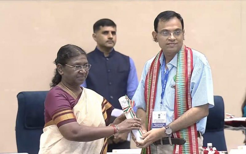
Teaching is much more than just imparting knowledge, it is an inspiring change. Learning is more than absorbing facts, it is acquiring understanding. When we recall our own education, we remember the teachers not methods and techniques. The belief of a teacher on his/her student can make them achieve wonders. Teachers are the root of an education system and can change lives with just the right mix of chalk and challenges. Teaching is the profession that teaches us all the other professions. On this momentous occasion of Teachers Day 2023, Prof. Suman Chakraborty, a professor in the Mechanical Engineering Department at the IIT Kharagpur has been selected for the National Awards to Teachers 2023.

If you can simplify the technology, you don’t need a doctor or a highly qualified technician to work on the technology; even an Asha Health Worker or a similar front line health worker can use it with minimal training,’ said Prof. Suman Chakraborty, a professor in the Mechanical Engineering Department at the IIT Kharagpur, who has been selected for the National Awards to Teachers 2023 by the Ministry of Education, Department of Higher Education, Government of India.

Your research has been on developing devices that would help rural healthcare. Is there any particular incident that caught your attention to this often neglected area?

If you look at the journey of any researcher, you will see there are certain things which happen by chance, rather without much planning. You develop certain insights from experience and that direct you to one particular direction. That is the case with me too. I worked on micro fluidics and its applications on healthcare, but not particularly focusing on rural healthcare. Though at IIT Kharagpur where I work has a township, it is basically located in a rural area. There is a railway station here and if you go beyond the railway station, the scenario is totally different. It is like any other rural area you see in India. It is difficult not to notice the huge contrast and difference between the kind of facilities and access to facilities we have at IIT and a place that is just a kilometre away from IIT. People there do not have either affordability or accessibility to healthcare, or a combination of these two.

It was a disturbing scenario…

Definitely. It still is disturbing. The focus of our work has always been on medical diagnostics, but when I noticed this disturbing disparity, we shifted our focus to the under-served sections of our society. That means, the technologies we use now can be used in the field and not in labs where you have air-conditioning, refrigeration and high-end machines. We have tried to solve the problem in some way, but we are yet to come out with a robust solution that will solve the problem completely. Yes, we could solve it in such a way that it is better than what it was earlier. If you look at the technological developments in healthcare, they are done with the assumption that everybody can afford it. You have these high-end labs and diagnostic centres manned by qualified technicians and doctors in big hospitals which are not accessible to everyone. Even if they are accessible, not many can afford them.

Reports say 80% of doctors in India work in urban areas. Do you think technology alone can solve a lot of healthcare deficiencies in rural areas?

Technology alone cannot solve the problem entirely, but most of it. If you can simplify the technology, you don’t need a doctor or a highly qualified technician to work on the technology; even an Asha health worker or a similar front line health worker can use it with minimal training.

Does that mean you were simplifying the technology so that they can use it in the rural areas?

Yes. That’s where the challenge comes; simplifying the technology without compromising on the performance. When you simplify technology, there is always a danger of compromising on accuracy. So, what we were developing had to be simple yet accurate and also cost-effective. Initially, you cannot go for simplification of technology as as you may miss some scientific components. It is a two-step process for us. First, we develop the technology in the lab so that we will know the nitty-gritties of the technology. Then, we move on to simplifying it.

Can you give us an example? For example, you have developed a device to check the haemoglobin level of individuals in the field and not in the lab…

There is a classical test used in all the labs which can be manual, semi-automated or fully automated. What we did was, we tried to understand the principle and then implement the principle on a piece of paper. We use just one drop of blood from the finger onto to a strip of paper and get the result within a minute. The amount of reagent we use also is very less, thus reducing the cost. What we do is, design the paper in such a way that the same principle we use in labs works on the paper too, that is the same chemistry and same reaction.

Is it an ordinary paper?

It’s a filter paper. When you compare the results of the test done on the paper and also in a lab, you will see that the results are more or less the same.

Is it for the initial diagnosis that these kits are used?

You can treat it as good as any standard lab test. The next step after the test is, take a picture of the colour of the blood to check the level of haemoglobin. For that, we keep the strip in a box, and with the help of a smart phone camera, the picture of the image is taken. Then the analysis of the colour of the blood is done by the app that is formatted in the phone. The image analysis and interpretation is done automatically by the phone and the person taking the test doesn’t have to do anything. Remember we do not expect the person, taking the test, for example as Asha worker to know anything about the technology. When the result comes on the app interface, it will be accessible to any doctor who is sitting in a big city. Like you said, 80% of the doctors are located in the urban areas. After looking into the test result and the patient’s history, the doctor can immediately give the first level of recommendation. For example, if the haemoglobin level is extremely low, and the patient requires an immediate blood transfusion, the caregiver can take him to a place where the patient can be administered blood. If it is only mild anaemia, the doctor may prescribe medicine or dietary changes.

Prof. Suman Chakraborty and researchers at IIT-Kharagpur are developing a technology to source electricity from clothes drying in open space

Was this the first device you developed?

The first one was to test the glucose level in the blood. The glucometer that is generally used by people is quite expensive for a person living in the rural area. We also developed another device to check the creatinine level as kidney is the first organ that gets affected by uncontrolled diabetes. Then, we have a device to check the lipid profile. We have this belief that only people in the urban areas suffer from high cholesterol. No, those in the rural areas also are affected. The diseases which we previously thought to be associated with urbanisation are there in the rural areas also because of the life style changes. Diabetes affects rural India more because they lack facilities to check the sugar level periodically. The idea behind all our inventions are, there is a need for early detection of diseases.

Do you use the same paper strip for all the tests, for example for haemoglobin, creatinine and glucose?

The paper is the same but we use different reagents for different tests.

Can we say almost all the common diseases are tackled at the preliminary stage through your devices?

Yes, you can say that. We have devices for these basic tests. Then we also have devices to diagnose infectious diseases like flu with which we can diagnose TB also which is a major disease in rural India. We use molecular diagnostics for infectious diseases which is one of the most difficult diagnostic technologies. But we have created a portable device which is an alternative to the RT-PCR machine. This machine we developed during the covid time is named COVIRAP. This machine is a 1ft x 1ft x 1ft box and it can perform the test like an RT-PCR machine, and the test results can be known within 40-45 minutes. Though we developed it for covid tests during the covid period, subsequently we use it influenza test. Now, we use it to detect TB. The advantage is, you can use the same device to do different tests by using different strips of paper with different reagents.

You have also developed a device to detect oral cancer…

If you notice, the device we spoke about earlier require body fluids to test. The device we developed to detect oral cancer does not require any body fluid; it is done through imaging. Though there are several reasons for oral cancer, majority of the patients are tobacco or gutka users, and they belong to the group who do not have access to early detection or care. If detected early, any ulceration in the mouth can be prevented from becoming cancer by changing their lifestyle. A large number of people could be saved if it is detected early. What we have developed is a device which looks like a torch with a thermal camera with which we can take the picture of any ulceration inside the mouth.The image we grab from inside the mouth is temperature at different points. The algorithm we have developed will convert the temperature into showing the blood flow in the area. If there is cancer in an area, new blood vessels grow (angiogenesis). There will be a significant change in the blood flow pattern in an area when there is cancer or pre-cancer.

You have used AI (Artificial Intelligence) in all your devices. How important is AI in developing devices like these?

AI is very important particularly in the healthcare space as there is a lot of difference between one person to another. Every human being is different. Health issues are also very personalised and you see variations in every person. In the normal conditions itself, even the physical appearance of say, the inside of the mouth of one person is different from another person. With the help of AI, we can predict the individual variations more accurately.

Do you consider what you are doing as part of your responsibility towards society?

Of course. Social responsibility can be addressed in different ways by different people. This is the way people working science and technology can impart their responsibility to society. What we are doing is not even 10% of what is needed. We have only introduced the technology; it has to be a part of the healthcare system and not present in an isolated manner here and there. And it cannot be done by the efforts of one or two individuals; it has to be a national movement and part of the entire system.

Some Major Awards & Accolades :

  • The Shanti Swarup Bhatnagar Award in 2013.
  • The J C Bose Fellowship in 2018.
  • The G D Birla Award in 2021.
  • The Infosys Prize in 2022.
  • National Award for Teachers 2023.

Media Coverage:

The Bengal Post Anandabazar Patrika 

Interview : Courtesy Rediff.Com

Edited By : Poulami Mondal, Digital & Creative Media Executive (Creative Writer)
Email: poulami.mondal@iitkgp.ac.in, media@iitkgp.ac.in, Ph. No.: +91-3222-282007

Follow us on: Facebook – IIT Kharagpur; Twitter – @IITKgp;  Instagram – @iit.kgp; LinkedIn – Indian Institute of Technology
For news visit: https://kgpchronicle.iitkgp.ac.in/

Digital CHAVI for Cancer Cure

Outlook   ET HealthWorld   Millennium Post   Lokmat   Financial Express   Business Standard    India Today    Amar Ujala    Navbharat Times   NDTV   New Kerala    The Quint    Indian Express    

IIT Kharagpur and Tata Medical Center have set up an open architecture image biobank to aid cancer research in the country. Named CompreHensive Digital ArchiVe of Cancer Imaging (CHAVI) it will address the emerging field of imaging-related research and will be India’s first step towards harnessing artificial intelligence and deep learning methods to answer medical questions of importance in the field of image banking.

IIT Kharagpur and Tata Medical Center have been jointly working on several novel educational and research programmes including Masters and Fellowship courses to enable this trans-disciplinary research that marries technology and medicine. The two institutions have joined hands in initiating a pilot project on developing an image data bank for cancer patients, in particular, the present focus is radio oncology. The project has been undertaken by IIT Kharagpur through the National Digital Library Initiative (NDLI) of MHRD. The overarching aim here is to build up a national bank of annotated images with a flexible query interface and link it with a pipeline of radiomic services for furthering radiomic research in large image datasets.

The CHAVI project is the first of its kind. The objective of the National Digital Library of India is to make accessible material for doing research that normally could not have been done in India. With the CHAVI project, as a beginning, we have chosen cancer imaging database along with Tata Medical Center because of their tremendous expertise. Cancer is one of the most dreaded diseases in our country. If we are able to create a very well defined, annotated database, it will help researchers as well as doctors to be able to do early, more accurate diagnosis and provide better treatment for our people which is a lot more cost effective – Prof. Partha Pratim Chakrabarti, Principal Investigator of NDLI.

As a pilot, radiation oncology related images are being banked within the NDLI CHAVI RO project. It is a prototype system which is under development addressing various such issues. It is also being developed considering multi-institutional participation in building a national image data bank.

Once the pilot project is successful, it can be scaled up to a larger set of medical images. Medical imagery can then be combined with AI to enable reach of treatment to more people as well as provide targeted therapy based on individual symptoms. This should enable doctors like never before, and revolutionize the way doctors interact with patients and systems.

AI for the medical vertical has three pillars. Descriptive analysis that will help education – students anywhere in the country can access the bank to look at the images and learn from there. Predictive Analysis will help doctors diagnose better. And then Prescriptive analysis that will help doctors reduce the scope of treatment based on past use cases.

We need more affordable solutions in India for cancer treatment, majority of our patients are middle class and lower middle class and cannot afford genomic analysis. Image banking combined with predictive/prescriptive AI can enable us to identify signatures as a much more cost effective alternative – Dr Sanjoy Chatterjee, Tata Medical Center.

While Tata Medical Center has created a large repository of medical data and images of cancer patients including outcomes of treatment in many cases, there are various challenges while building this system. The first and foremost is in preserving anonymity of patients as well as maintaining adequate referential integrity, a necessity for carrying out useful research.

To enhance the CHAVI project, the two institutions organized a workshop titled – “Structuring a Collaborative National Image Banking Program” on 26th July 2019 at Tata Medical Center, Kolkata, supported by MHRD through the NDLI project. The workshop which was coordinated by Dr. Sanjoy Chatterjee and Prof. Jayanta Mukhopadhyay from IIT Kharagpur involved presentations and panel discussions with experts in medical and Computer Science / AI domains. Several expert doctors from India, USA and UK and specialists in the area of Computer Science from India also took part in the daylong proceedings.

Who said what in the workshop

The scope of image banking is to enable cancer research and move it forward, to access data that is more diverse and come from different centres, different patients and different ethnic groups to help doctors make more informed decisions and deliver personalized treatments – Dr. Emiliano Spezi, Cardiff University, USA

For research, we need geographic distribution – which means we need to build national archives, be it central or distributed and then connect them globally to be truly able to sample the human population – Dr. Fred Prior, UAMS, USA

If you have an image bank where you can collaborate all your images, and then you look at certain features, you can probably come up with information which goes beyond the human eye. Imaging when combined with pathological information can then improve outcomes for our patients – Dr. Simon Pavamani, CMC Vellore

It is a kind of personalized medicine. Where a set of images of a particular kind is treated in a particular way which helps predict a specific treatment for each individual patients – Dr. Subhas Gupta, AIIMS, New Delhi

Smart Diagnostics for Pulmonary Healthcare

Business Standard (IANS)   Business Standard (PTI)    Deccan Chronicle   Biospectrum     Outlook    Quint     Devdiscourse

Researchers at the Dept. of Electronics and Electrical Communications Engineering at IIT Kharagpur have developed a decision support system to diagnose malignant and other diseased tissues in the lungs. While one system can refer to CT scan images to detect lung nodules and test them for the possibility of malignancy, a second software can detect Interstitial Lung Disease (ILD) patterns in chest HRCT images.

“Biopsy especially in the lungs is a critical process, hence conducted only after initial medical analysis is done by expert radiologists. The developed systems use noninvasive and comparatively affordable methods of image analysis that would aid the radiologists to identify malignancy by reading growth in the lung nodules. The other system will help identify interstitial disease patterns in HRCT images depicting the lung tissue texture,” explained lead researcher Prof. Sudipta Mukhopadhyay.

“The novelty of the system lies in its India-centric reference point i.e. the medical image scan database used for reference is sought from the Indian patient population. We worked with Prof. Khandelwal and his team from PGIMER Chandigarh for data ground truth and clinical data. Also, foreign database such as LIDC-IDRI and MedGIFT ILD database has been used. The biopsy cases were primarily taken from PGIMER,” explained researcher Shrikant Mehre.

The malignancy detecting tool detects the lung nodule, segment the nodule, and provides a way to modify segmentation, retrieve similar nodules from the database with their report and assess the chance of malignancy of the query nodule based on the retrieval results. The ILD tool is developed by incorporating feedback from expert radiologists to make it easy to use for non-tech savvy clinicians. The software is equipped with necessary modules such as automatic segmentation of lung boundary and pathological region within lung area, provision to modify the boundary, retrieving similar segments from the database with their report and assess the probability of the pathological segment to be a particular ILD category based on the retrieval results. The mapping of disease is performed by doctors based on the ILD pattern and clinical inputs.

“We have successfully tested both software systems at AIIMS Delhi. Prof. Ashu Seith Bhalla and her team provided the neutral test site required for the validation. Currently, lung nodule detection rate and classification rate is 86% and 87%, respectively, and the success rate for ILD classification is 84%. We are working towards further improvements in order to conduct clinical trials on bigger sample sizes,” said another researcher Mandar Kale.

With the growing cases of cancer and other respiratory diseases in India, the need for skilled radiologists is expected to grow exponentially in near future. Budding radiologists will be highly benefited in learning from previous images stored in Picture Archival and Communication System (PACS) and reports in Radiological Information System (RIS) on their own and to help practicing radiologists in differential diagnosis using the CBIR based Computer Aided Diagnosis (CAD) system.

The research has been reported in more than 13 international journals and 19 international conferences through its various stages of progress.