Pandemic Healthcare Technologies Underway @IITKGP

Economic Times          Times of India            Times of India (PTI)            Hindustan Times            Outlook          Indian Express       CNN News18     News18 Bangla (TV)           News18 Bangla (Web)         Deccan Herald         Careers360          Analytics India Magazine          NDTV            Firstpost           India Blooms           Business Insider        The Week          Aaj Tak         India Today

IIT Kharagpur has set up research funding for R&D work related to COVID-19. The Institute submitted a list of projects to the IIT Council last week of which 8 projects have been selected.

Dr Ramesh Pokhriyal Nishank, Hon’ble Minister, Ministry of Human Resource Development, Govt. of India appreciated and acknowledged the initiative by the Institute on his social media handle congratulating the Director Prof. Virendra Kumar Tewari and his team of researchers.

Talking about the initiative Director Tewari said “It is our responsibility to improve the quality of life of the last person in the society. While we built some quick technologies to cater to the immediate needs of the essential service providers at the campus, we were simultaneously preparing project proposals and evaluating them keeping in mind the immediate need of the country, cost and product delivery period.”

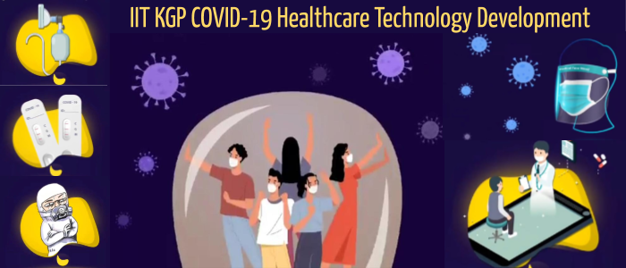
The researchers would be working on several technologies including design and development of rapid diagnostic kit, real-time PCR machine, body suit for COVID-19 patients, personal protective equipment for healthcare workers and portable shredder integrated with sterilizer, Hazmat Suit with forced purified and cooled air circulation for medical professionals, bootstrapping ambu-bag as automated ventilator, telemedicine for fighting viral pandemic, large scale production of recombinant proteins for vaccine and testing.

An amount of Rs. 50 Lakh has been allotted for phase I of 8 projects towards development of prototypes. For most of these projects, the prototypes are expected to be ready within a duration of 3 – 4 weeks, while a couple of them would take about 6 months to deliver the results. The phase I is expected to start immediately after the lockdown is relaxed and the research staff are able to attend the laboratories. Meanwhile software related work would progress as usual.

“IIT Kharagpur has a proven track record towards development of indigenous health and hygiene technologies which are affordable, high-quality at par with globally accepted standards, and commercially viable. Our researchers are committed to deliver the prototypes within a constrained timeline considering the healthcare needs in the current situation,” added Prof. Tewari.

1 Development of smartphone-integrated paper-strip kit for rapid low-cost diagnostics of COVID-19 infection Prof. Arindam Mondal and Prof. Suman Chakraborty
2 Design and Development of an indigenous Real Time PCR Machine Prof. Anandaroop Bhattacharya, Prof. Prasanta K. Das, Prof. Suman Chakraborty (ME Dept), with inputs from Dept. of Biotechnology and Physics
3 Towards large scale Production of Recombinant Proteins for Vaccine and Testing of Novel COVID-19 Prof. Sudip K. Ghosh, Prof. Ananta K. Ghosh and Prof. Ramkrishna Sen
4 Bootstrapping the ambu-bag as automated ventilator Prof. Aditya Bandopadhyay + Faculty and Students from ME Dept
5 Design and Development of a Bodysuit for COVID-19 Patients to Prevent the Spread of Infection Prof. Nishant Chakravorty
6 Telemedicine for fighting viral pandemic such as COVID-19 Prof. Jayanta Mukhopadhyay
7 A Hazmat Suit with Forced Purified and Cooled Air Circulation for Medical Professionals Prof. Manoj Kumar Mondal
8 Personal Protective Equipment for Health Care Workers
Prof. Santanu Dhara and Prof. Sangeeta Das Bhattacharya
8.a. Portable shredder integrated with sterilizer

SPARC Workshop Explores Critical Challenges of Children’s Healthcare

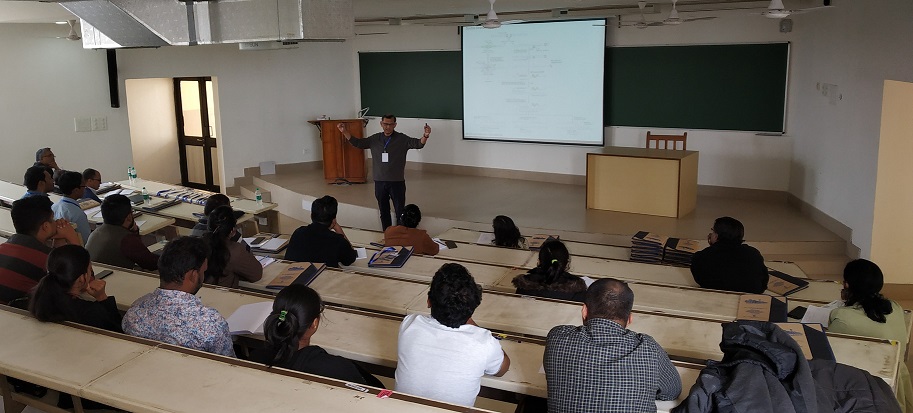
The School of Medical Science and Technology at IIT Kharagpur recently held an Indo-UK Residential Workshop in collaboration with the University of Manchester, UK, on the “Practical Management of Inherited Pediatric Hematological Disorders”. The focus was on clinical reasoning of cases related to children’s blood and bone marrow disorders using an interactive problem-based learning approach with direct interactions with globally renowned experts.

The areas covered in the workshop included the workup of the child presenting with signs and symptoms of reduction in the number of various blood cells, or cytopenias including pancytopenia or bone marrow failure. Workup included understanding the genetics behind certain syndromes, targeted testing, and treatment planning including an overview of bone marrow transplantation. Other topics covered included understanding cognitive errors in clinical decision making and computerized clinical decision support systems.

Experts from IIT Kharagpur, University of Manchester, Tata Medical Center and Tata Translational Cancer Research Centre, Kolkata, and NRS Medical College participated in this three-day workshop which was sponsored under the Scheme for Promotion of Academic and Research Collaboration (SPARC), an initiative of Ministry of Human Resource Development, Govt. of India.

Among the speakers were Prof. Vaskar Saha from the University of Manchester and Director of the Tata Translational Cancer Research Center a pediatric oncologist and hematologist and an expert in the care of children with leukemia. He worked through cases with participants to guide thinking on how to approach the child with a complex bone marrow disorder to come up with a diagnosis that could then lead to a treatment plan. Dr. Shekhar Krishnan, senior consultant in paediatrics hematology and oncology at TMC-TTCRC discussed bone marrow transplant.

Diagnosis was a focus area of the workshop. Dr. Niharendu Ghara senior paediatric consultant at TMC-TTCRC Kolkata discussed targeted approaches to diagnostic testing making sure to find the right test for the right situation, Dr. Rajib De from NRS Medical College discussed thalassemia in the context of Eastern India and approaches to screening, diagnosis, and management.

Prof. Jayanta Mukhopadhyay from IIT Kharagpur’s Dept. of Computer Science and Engineering discussed automated clinical decision support systems and their role in improving clinical decision making. Prof. Sangeeta Das Bhattacharya of SMST discussed cognitive errors in clinical decision making.

Students from diverse areas such as MBBS, Masters in Medical Science & Technology, MD, postdoctoral fellows, and clinical fellows in hematology participated in the workshop. The participants gained insights from master clinicians in real-time in clinical reasoning through case-based learning.

Digital CHAVI for Cancer Cure

Outlook   ET HealthWorld   Millennium Post   Lokmat   Financial Express   Business Standard    India Today    Amar Ujala    Navbharat Times   NDTV   New Kerala    The Quint    Indian Express    

IIT Kharagpur and Tata Medical Center have set up an open architecture image biobank to aid cancer research in the country. Named CompreHensive Digital ArchiVe of Cancer Imaging (CHAVI) it will address the emerging field of imaging-related research and will be India’s first step towards harnessing artificial intelligence and deep learning methods to answer medical questions of importance in the field of image banking.

IIT Kharagpur and Tata Medical Center have been jointly working on several novel educational and research programmes including Masters and Fellowship courses to enable this trans-disciplinary research that marries technology and medicine. The two institutions have joined hands in initiating a pilot project on developing an image data bank for cancer patients, in particular, the present focus is radio oncology. The project has been undertaken by IIT Kharagpur through the National Digital Library Initiative (NDLI) of MHRD. The overarching aim here is to build up a national bank of annotated images with a flexible query interface and link it with a pipeline of radiomic services for furthering radiomic research in large image datasets.

The CHAVI project is the first of its kind. The objective of the National Digital Library of India is to make accessible material for doing research that normally could not have been done in India. With the CHAVI project, as a beginning, we have chosen cancer imaging database along with Tata Medical Center because of their tremendous expertise. Cancer is one of the most dreaded diseases in our country. If we are able to create a very well defined, annotated database, it will help researchers as well as doctors to be able to do early, more accurate diagnosis and provide better treatment for our people which is a lot more cost effective – Prof. Partha Pratim Chakrabarti, Principal Investigator of NDLI.

As a pilot, radiation oncology related images are being banked within the NDLI CHAVI RO project. It is a prototype system which is under development addressing various such issues. It is also being developed considering multi-institutional participation in building a national image data bank.

Once the pilot project is successful, it can be scaled up to a larger set of medical images. Medical imagery can then be combined with AI to enable reach of treatment to more people as well as provide targeted therapy based on individual symptoms. This should enable doctors like never before, and revolutionize the way doctors interact with patients and systems.

AI for the medical vertical has three pillars. Descriptive analysis that will help education – students anywhere in the country can access the bank to look at the images and learn from there. Predictive Analysis will help doctors diagnose better. And then Prescriptive analysis that will help doctors reduce the scope of treatment based on past use cases.

We need more affordable solutions in India for cancer treatment, majority of our patients are middle class and lower middle class and cannot afford genomic analysis. Image banking combined with predictive/prescriptive AI can enable us to identify signatures as a much more cost effective alternative – Dr Sanjoy Chatterjee, Tata Medical Center.

While Tata Medical Center has created a large repository of medical data and images of cancer patients including outcomes of treatment in many cases, there are various challenges while building this system. The first and foremost is in preserving anonymity of patients as well as maintaining adequate referential integrity, a necessity for carrying out useful research.

To enhance the CHAVI project, the two institutions organized a workshop titled – “Structuring a Collaborative National Image Banking Program” on 26th July 2019 at Tata Medical Center, Kolkata, supported by MHRD through the NDLI project. The workshop which was coordinated by Dr. Sanjoy Chatterjee and Prof. Jayanta Mukhopadhyay from IIT Kharagpur involved presentations and panel discussions with experts in medical and Computer Science / AI domains. Several expert doctors from India, USA and UK and specialists in the area of Computer Science from India also took part in the daylong proceedings.

Who said what in the workshop

The scope of image banking is to enable cancer research and move it forward, to access data that is more diverse and come from different centres, different patients and different ethnic groups to help doctors make more informed decisions and deliver personalized treatments – Dr. Emiliano Spezi, Cardiff University, USA

For research, we need geographic distribution – which means we need to build national archives, be it central or distributed and then connect them globally to be truly able to sample the human population – Dr. Fred Prior, UAMS, USA

If you have an image bank where you can collaborate all your images, and then you look at certain features, you can probably come up with information which goes beyond the human eye. Imaging when combined with pathological information can then improve outcomes for our patients – Dr. Simon Pavamani, CMC Vellore

It is a kind of personalized medicine. Where a set of images of a particular kind is treated in a particular way which helps predict a specific treatment for each individual patients – Dr. Subhas Gupta, AIIMS, New Delhi

Sights and sounds


India Education Diary

Medical imaging is a multi-billion-dollar industry today and is expected to grow at a CAGR of 5.7% by 2025. Both India and China are expected to majorly contribute to this growth. A rapid advancement in medical and diagnostic imaging devices has enhanced the diagnosis and treatment of several diseases, including cardiovascular diseases and cancer.

In addition, medical imaging and image guided therapy are important sources of employment, with a large number of electrical, computer, and biomedical engineers being employed in both the public and private sectors. However, today there are only a handful of academic programs (mostly in medical imaging or medical physics) that focus on training students to enter these fields.

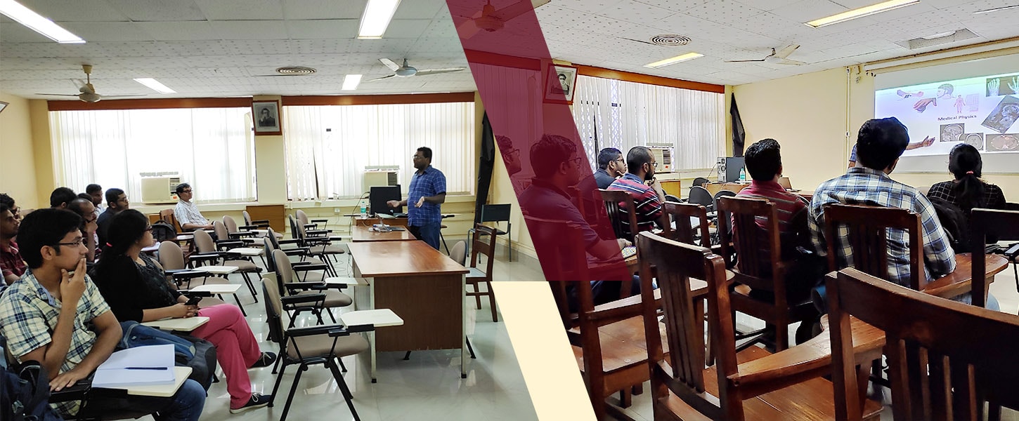
An ongoing short term course at IIT Kharagpur addresses this problem by not only trying to keep students abreast of the latest trends in the imaging sciences but also in helping generate an interest in this particular field. Dr. Soumyajit Mandal, T. and A. Schroeder Assistant Professor from the Department of Electrical Engineering and Computer Science of Case Western Reserve University , is currently taking the week-long course on “Instrumentation and algorithms for biomedical imaging: MRI and Ultrasound”. The course is aligned towards instrumentation and signal processing algorithms (or strategies) while focusing on physics and engineering of the applicable methods.

Dr Mandal said at the inaugural lecture, “The typical course on imaging methods deals either with the physics of the problem (how images are created or what is the physical phenomenon involved) or the clinical aspects. I will talk about the engineering of the system, say what sort of circuits are used, and such like.”

Dr Mandal is a recognized expert in the area of bioinspired electronic circuits and medical imaging. He is an alumnus of IIT Kharagpur, and won the President’s Gold Medal when he graduated in 2002. His doctoral thesis at MIT won him the MTL Doctoral Dissertation Award in 2009. Last year, he was awarded the Young Achiever Alumni Award by his alma mater.

The course is being hosted under the Institute’s SGR International faculty/expert outreach program that funds distinguished researchers or faculty members of highly-ranked international universities to teach and do collaborative research in IIT Kharagpur. Since its launch in 2015, SGRIP (Shri Gopal Rajgarhia International Programme) has facilitated the visits of more than 40 international faculty and led to about 25 collaborative short courses and workshops at IIT Kharagpur.

Prof. Anandaroop Bhattacharya, Associate Dean, International Relations, said, ““The SGRIP program, set up with the generous funding received from our Distinguished Alumnus, Mr. Shri Gopal Rajgarhia, aims at bringing eminent researchers across the world to IIT Kharagpur to share their research work with our students and faculty. The initiative goes a long way in stimulating the learning environment and promoting collaborative research. This particular course work exposes our students to cutting-edge research in one of the emerging fields of science.”

Prof. Sudip Nag of the Department of Electronics and Electrical Communications Engineering, who facilitated the course being taken by Dr Mandal, pointed out, “This course is an awesome platform to learn about basic medical imaging physics, smart circuit design approaches, and futuristic signal processing strategies. This course has been expanded through magnetic resonance imaging (MRI) and ultrasound imaging as the prime thematic areas, while simultaneously dealing with the latest and upcoming trends in portable healthcare image acquisition, reconstruction methods, and machine learning integration in healthcare. The course will certainly and immensely benefit students, researchers, and faculties at IIT Kharagpur who are directly or partially engaged in related areas of research.”

The course is being attended mostly by senior students doing their MTech or PhD. Baisakhee Saha, a woman scientist associated with the Institute’s School of Medical Science and Technology, who is attending the course said, “I am interested in micro-CT imagery and keen to know about the advance of cellular imagery. I am looking forward to the course for new directions.”